The hospital is equipped with all the disciplines/ modalities required for full and proper diagnosis of cancers. The facilities of Pathology , Radiodiagnosis & Imaging and Nuclear Medicine are open to use for non-cancer patients also. As cancer is a non -communicable disease, there should be no hitch in utilizing these facilities for the diagnosis of various other diseases.
Diagnostic Facilities
PATHOLOGY
Department of Pathology is one of the most important departments for a cancer hospital, as correct diagnosis depends upon accuracy of various pathological tests and competence of pathologist. All pathological diagnostic modalities required for diagnosis of cancer as well as diagnosis of other ailments are available in this broad- based set up.
Fully equipped laboratories with modern sophisticated and automated gadgets are used for various procedures. The Department of Pathology has facilities for Histopathology, Cytology ,Microbiology, Biochemistry, Haematology, Tumour Markers, Electrophoresis, Blood Bank etc. Well-qualified and trained pathologistis and technicians are looking after the work of this vital wing of the institute.
(I) Clinical Biochemistry
The department is doing routine biochemistry tests like, Liver function tests, Renal function tests, Lipid Profile etc. on a Semi Automatic Biochemistry Analyzer machine (Transasia), and Electrolyte estimation using Automatic Electrolyte Analyzer (Transasia).
(II) Hematology
All basic and specialized hematological investigations especially required for diagnosis of hematological and lymphoid malignancy like Bone Marrow Aspiration, Trephine biopsy and Cytochemistry besides routine CBP and Complete Haemogram are performed by skilled hands.
(III) Cytology
Fine Needle Aspiration Cytology (FNAC) plays a very important role in diagnosis of all type of lesions. It is a very simple, painless, cost effective and quick, outdoor procedure for which no anesthesia is required.
(IV) Histopathology
Histopathology is a gold standard for the final diagnosis. There are very good quality Microtomes, Automatic Tissue Processor (Shandon, UK) and other modern equipments required for quick tissue processing. We also have Cryostat (Shandon UK) for intraoperative diagnosis.
(V) Tumour Markers
We are routinely doing FT4, TSH, CA-125, CA 15-3 CEA, tPSA, β-HCG, AFP and IgM, IgG, antibodies for Tuberculosis on Elisa Strip Reader (Merck).
(VI) Immunochemistry
This is a very important investigation for the final diagnosis of difficult and unusual cases. Besides being of routine use in breast cancer for prognosticating the patient, it also helps to choose specific treatment modalities. At present , we are performing ER, PR, Her-2 Neu Pancytokeratin, HMWCK, EMA, CEA, Vimentin , CD -117, LCA, CD-3, CD-20,CD-15, CD-30, GFAP , Thyroglobulin, Calcitonin , NSE , Chromogranin A, HMB-45, MIC-2,AFP, HHF-35, ,Desmin and S-100.
(VII) Blood Bank
The institution is also having its own Blood Bank, which caters to the need of patients under a very efficient and well-organized system. We will be starting blood component preparation in near future.
The well equipped Radiodiagnosis and imaging department is the backbone of any modern hospital and treatment centre in present era.
The quality of treatment which any good hospital can give to the patients depends on excellence of the diagnostic and imaging department of the hospital. All the faculties of hospital , in some way or other, inevitably take the help of radiodiagnosis and imaging in order to reach the proper diagnosis so that correct treatment can be planned.
It is departments endeavour to help all the specialists in their fields to diagnose the various types of diseases with the help of conventional X-Ray, USG, Colour Doppler Mammography and CT Scan and to locate the disease and assess the extent. Interventional procedures like USG and CT guided FNAC, biopsies are also done to reach a cytological and histological diagnosis. Therapeutic aspirations are also performed under ultrasound and CT guidance.
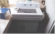
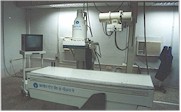
The department has one 500mA X-Ray machine with Image Intensifier that function round the clock to carrying out all types of plain X rays and special investigations like Barium studies of GIT,IVP,MCU and RGU for radiography requirement of out patients and inpatients. Two portable X ray Units are available for routine radiography of the immobile inpatients
500 MA X-Ray Machine With Image Intensifier
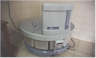
Ultrasound Unit with
Colour Doppler LOGIQ-5 Pro
| Highly sophisticated LOGIQ-5 Pro Colour Doppler and Ultrasonography unit with four Transducers (Convex, Linear, Cardiac, and Transrectal/Vaginal ) is available for carrying out sonography of whole abdomen ,high resolution sonogram of small parts like breast, neck, orbit, scrotum, thyroid and musculoskeletal system. Transrectal and Transvaginal sonogram are also performed. Entire range of color Doppler studies including neck vessels , visceral and peripheral blood vessels are being performed to assess the abnormalities of blood vessels. |
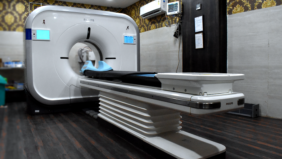
High-Quality Multi-Detector CT Machines: A 128-Slice And A 16-Slice MDCD
The Radiology Department offers comprehensive services encompassing all aspects of Radiological Imaging and Image-guided non-vascular Interventional Procedures. Our commitment to early detection is evident in our specialized screening mammogram for Breast Cancer.
Equipped with state-of-the-art technology, our facility boasts two high-quality multi-detector CT Machines: a 128-Slice and a 16-Slice MDCD, catering to both Oncology and Non-Oncology CT studies. Our ultrasound capabilities are extensive, featuring multiple machines with Color Doppler functionality and various frequency probes. We provide portable ultrasound machines for bedside and operating room use.
X-ray services include a 600 MA machine with IITV for x-rays and related procedures, along with portable x-ray machines for bedside applications. Our department houses a cutting-edge Multi-detector CT Scan (MDCT) – 16 slice – SOMATOM Emotion, performing a diverse range of diagnostic studies.
In addition to whole-body CT imaging, we specialize in dual-phase CT scans of the liver, pancreas, and vascular studies such as spiral CT angiography for cerebral, carotid, abdominal, thoracic, and peripheral vessels. Using a pressure injector, we achieve accurate vessel depiction without invasive procedures. Virtual colonoscopy, endoscopy, and laryngoscopy are integral components of our service offerings.
Routine procedures include high-resolution CT studies for lungs and assessments of bony abnormalities like the temporal bone. We employ 3D reconstruction techniques, forming MPR and SSD from axial images when necessary. Ensuring image quality is paramount, we administer an adequate quantity of non-ionic contrast media to prevent adverse reactions. At our Radiology Department, we prioritize precision, advanced technology, and patient well-being in every diagnostic and interventional procedure we undertake.
Interventional procedures including diagnostic procedures like USG or CT Guided aspiration/ Biopsy are being performed routinely. Transvaginal biopsy of pelvic masses and transrectal biopsy of prostate are also performed.
Therapeutic procedures like drainage of fluid collections or abscess and celiac plexus block are being performed.
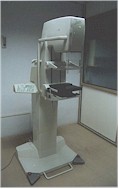
| Department is equipped with the latest imaging technology Planmed Sophie Classic Mammography unit – imported from Finland. It is highly advanced and sophisticated unit available in Madhya Pradesh. For basic screening mammography or for more sophisticated diagnostic procedures Planmed Sophie unit is ideal configuration. It provides quality images with low dose radiation exposure. It captures the maximum amount of breast tissue on the image , maximizing the field of view and enabling earliest detection of breast cancer. Ever since its installation, it has been of great help in early diagnosis and screening of the breast cancer in this region |
MRI facilities are provided in collaboration with leading centres of the city under smooth and prompt arrangement.
Department of radio diagnosis and imaging is managed by highly experienced and qualified team of radiologists and technicians.
Nuclear Medicine is a speciality where radioactive materials are used for diagnostic and therapeutic purposes. Diagnostic scans are physiological and non invasive.
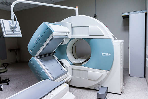
Nuclear Medicine is another speciality which is very essential for early diagnosis and treatment of various disorders. The department of Nuclear Medicine is equipped with the most modern e-cam Gamma Camera with SPECT facility. This is a very sophisticated equipment. The hospital has been the first medical institute in the country to procure this rare equipment. It is extremely useful for the patients who need Nuclear Medicine diagnostic imaging in Oncology (Whole Body Bone Scan), Cardiology (Thallium Test), Nephrology (Renal Scan), Endocrine disorders (Thyroid diseases), Gastroenterology (Liver Scan) and various Orthopaedic (Bone diseases) problems. It is also equally useful for non-cancer patients. This is the only nuclear medicine center in the states of Madhya Pradesh and Chhatisgarh offering Radio Iodine Therapy for Thyroid Cancer patients.
Evaluation of orthopaedic problems e.g. Trauma, Stress or Occult fracture, Osteomyelitis, Bone tumours, Paget’s disease, Assessment of viability of bone graft.
Urogenital system: Assessment of Renal functions, Congenital anomalies, GFR estimation, Renovascular hypertension. Assessment of Renal Morphology, Scars in Pyelonephritis, Renal Tumours, Vesiscoureteric reflux & Follow up of Renal Transplantation.
Gastroenterolog: Diagnosis of Acute Cholescystitis, Cirrhosis, Buddchiari Syndrome, Nodular hyperplasia, Tumour, Haemangioma, Detection of GI bleeding, Meckel’s Diverticulum. Assessment of GB dysfunction, Atresia and post operative cases. GE reflux assessment.
Endocrinology: Detection of Thyroid and Para Thyroid Disorders, Diagnosis of Pheochromocytomas and other non endocrine tumours, Treatment of Thyrotoxicosis and Thyroid neoplasm
Cardiolog: Myocardial perfusion scan to evaluate chest pain, To diagnose coronary artery disease. Assessment of severity and extent of Ischemia of Cardiac muscle and risk of infarction. Myocardium assessment before and after by pass surgery. Assessment of Myocardial viability in segments with Wall Motion abnormality. Estimation of LV functions and ejection fraction of IHD.
Respiratory system: Diagnosis of Pulmonary Embolism. Lung Cancer and Metastatic disease in the lungs. Cerebrovascular system Assessment of Stroke. To detect Epileptic focus and To evaluate dementia, Amnesia, Alzemier’s disease.
Brain Imaging: For Space Occupying Lesions, Infarct Vascular Malformations etc.
Diagnosis of deep vein thrombosis: Can be done with the help of Gamma Camera Images.
Whole Body Bone Scanning: For staging of Cancer, extent/spread of disease, Treatment of metastatic lesions and follow up of cancer patients.
In this context it would be appropriate to point out that on 16th March, 2002 Dr. B.A. Krishna, Head of the Department of Nuclear Medicine, PD Hinduja Hospital, Mumbai delivered a very illuminating lecture on clinical application of Nuclear Medicine. He opined that e-cam Gamma Camera is essentially a Nuclear Medicine Device designed specially for various imaging and scanning in regard to disorders and alterations pertaining to Breast, Liver, Lung, Brain, Thyroid, Kidney, Bone & other tissues. Such imaging is required in complex cases of cancer and in various conditions in Cardiology, Neurology, Endocrinology etc. It is not only a very reliable and useful diagnostic device required for cancer treatment but also equally useful and beneficial for scanning of wide range of other diseases, affecting different systems in body.
The department is also having the following facilities :-
- Thyroid probe : for thyroid uptake study.
- Stress lab for cardiac study: Thallium -201, 99MTC-Sestamibi and Tetrofosminis used for perfusion scan to assess cardiac status. This is very essential before any type of surgical intervention like bypass surgery or angioplasty etc. is done and for follow-up.
- Radio-immuno assay laboratory: Well equipped lab with well counter is available for various types of invitro studies including tumours markers.
- Radioactive Iodine therapy ward : Two rooms for the purpose have been made as per guidelines of Bhabha Atomic Research Centre for therapy in cancer thyroid and other types of malignancies. This is the first and only centre in Madhya Pradesh and central India for radioactive Iodine therapy facility.
- Gamma Surgical Probe : This is second such instrument available in India. This special device by which one can delineate tumour and lymphnodes through the use of specific radio pharmaceuticals with the help of gamma probe. This helps the surgeons to remove the tumour and lymphnodes with little morbidity used in Breast Conservation Surgery, Skin Cancer, Prostate Cancer etc.
- Research Activities:Student of M.D., M. Sc.(Biotechnology) and M. Pharma have done their thesis and research activities in the department.
According to eminent experts Dr. G.N.Mahapatra, Senior Consultant of Madakini Nuclear Imaging Centre, Mumbai and Dr.A.Malhotra Head of Nuclear Medicine department ,AIIMS New Delhi. the Nuclear Medicine Department of the hospital is one of the best equipped and organized department in the entire country.
All the diagnostic facilities are open to use by non cancer patients. As cancer is a non communicable disease patients suffering from other diseases should not hesitate in availing these facilities. The reports are provided on the same day of study.
Jawaharlal Nehru Cancer Hospital & Research Centre Bhopal
Refund & Cancellation Policy | Terms & Conditions | Privacy & Policy | © 2021. JNCHRC. All rights reserved. Averoft